
COTW 11/13/23: 53 yo F with elbow pain
This week we are talking about MSK ultrasound!
This week we had a lady that came in to our ED with left elbow pain after a fall while walking her dog. She tripped and fell unable to break the fall and fell directly on her left elbow. Initially she was feeling ok but the elbow pain persisted and she decided to come to get her elbow checked.
Apparently dog walking is now an extreme sport!
At first the left elbow was swollen compared to the right one. And so we decided to use the ultrasound to evaluate for any deviations from normal. Now how does a normal elbow ultrasound look like? you may ask. Here is an example:
Normal elbow long view
Normal elbow short view
These are normal views of the posterior aspect of the elbow showing the olecranon and the olecranon fossa. This is a great approach to evaluate for joint effusions, bone discontinuities along the distal humerus, triceps tendon tears or muscle tears, among others. You may want to check our COTW 02/24/23 that discussed the elbow anatomy as well.
Now that you have seen what normal looks like, let’s have a look at this lady’s elbow.
Pathologic elbow long view
Pathologic elbow short view
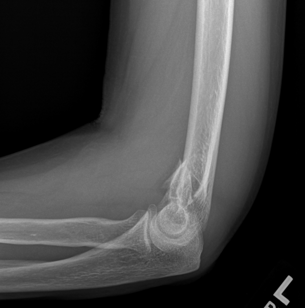
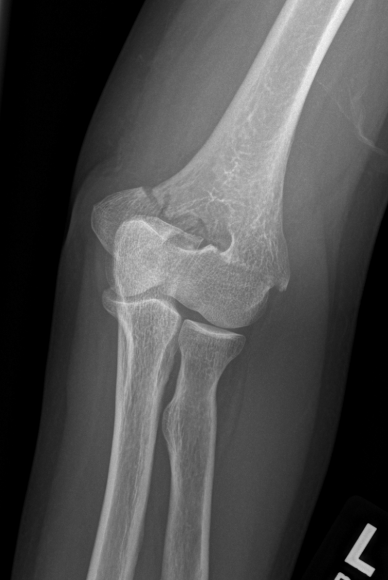
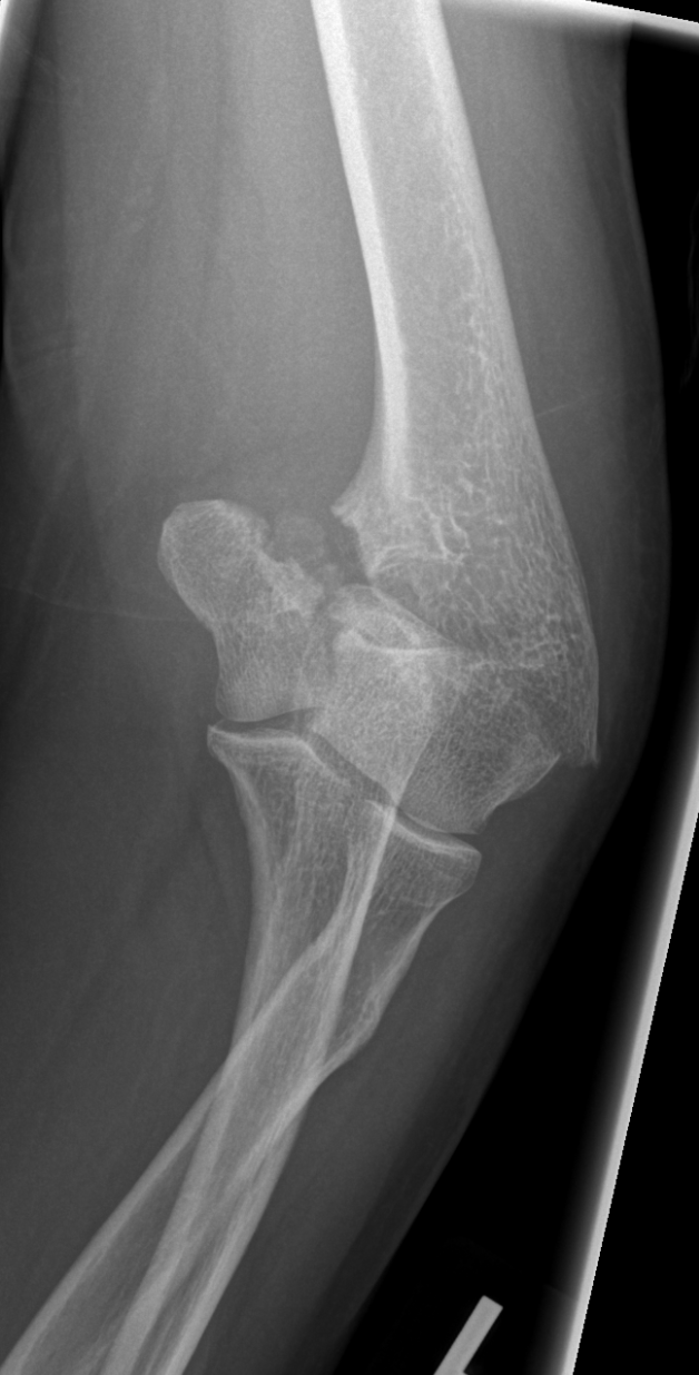
As you can see in these images there appears to be a difference in the relationship between the olecranon and the olecranon fossa. It seems deeper and you cannot see the fossa completely. Although it looks weird we did not notice any joint effusion. As we continued to evaluate the joint by fanning and swiping around the joint we noticed that there are discontinuities on the humeral bone cortex, something just doesn’t seem right, this “this sounds like a supracondylar fracture” we said (no pun intended). So we decided to the x-rays to confirm our ultrasound diagnosis.
After seeing all of this ugliness we decided to do a CT scan. Here is an image of the 3D rendering of the scan.
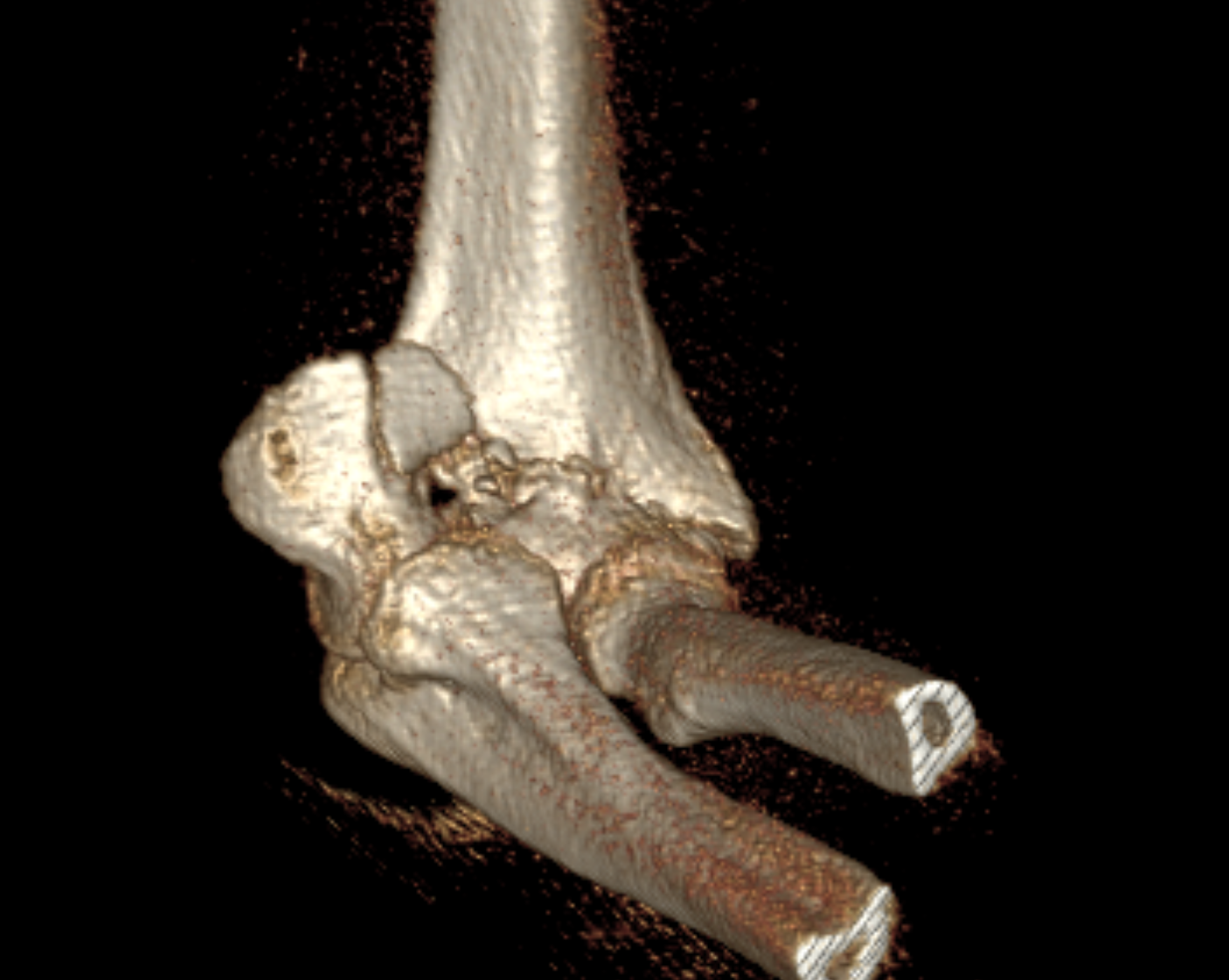
This patient was referred to orthopedic surgery for surgery.
The point of this case is to show you that MSK ultrasound may not provide the final diagnosis but it can give you a clear idea of what you are working with since your first contact with the patient. The more scans you do, the more familiarized you will be with the normal anatomy, remember all of what the body has two of has a control you can compare.
Just remember, if you have gel, you will scan.