
COTW 4/6/23: 48F finger pain and swelling
48 yo female presenting with 3-4 weeks of worsening L middle finger pain and swelling. Developed a mucoid dermal cyst on the dorsal aspect of her DIP joint that had intermittent scant serous drainage throughout the last few weeks. Swelling and pain have progressed especially in the last week and is now diffuse throughout her finger. Pain worse with both flexion and extension as well as palpation, and is worse on the palmar and lateral aspects of her finger. Denies trauma to her hand, fevers, red streaking up her hand.
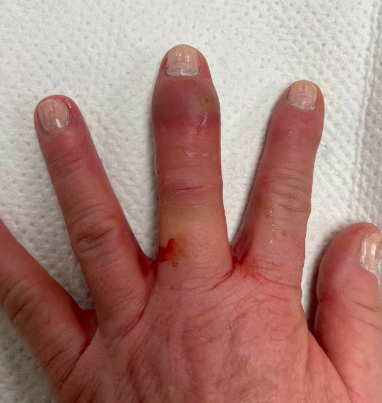
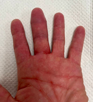
It’s somewhat hard to appreciate on these photos but the finger is more erythematous and slightly ecchymotic compared to the others. The finger isn’t particularly warm compared to the others. Patient has pain with flexion and extension but she states it is more on the sides of her finger. She has tenderness along the entire finger not just the FTS. Her finger is held in extension at rest.
An XR is done for you in triage. The final read comes back while you’re talking to the patient: Avulsion fracture of the third distal phalangeal base. The third distal interphalangeal joint and oblique nondisplaced fracture of the third digit middle phalangeal head.

You ask again and the patient swears up and down she hasn’t had any falls, jammed her finger, gotten hit, etc any time in the last few weeks to month.
Of course we’re worried about FTS… but it isn’t a perfect fit - she only meets 1-2/4 Kanaval criteria. Also in the differential are cellulitis, abscess, septic joint.. and now traumatic hematoma is back on the differential.
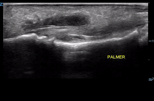
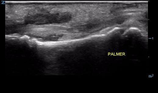
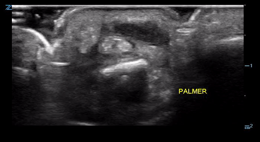
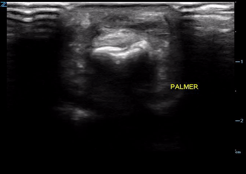
You can see the flexor tendon in the photos above as well as a separate fluid collection superficial to and distinct from the flexor tendon. While there is what appears to be an anechoic substance on both sides of the flexor tendon in the transverse view, you know that this isn’t swelling because it isn’t circumferential and disappears as you fan the probe and slide proximal. You remember this is just edge artifact.
You’re most concerned about a small abscess but with the underlying fractures this could could be a hematoma. With ortho at bedside you attempt to an US guided needle aspirate the abscess but don’t get much back even though you are clearly in the pocket.
Note needle tip in fluid collection in top left of image.
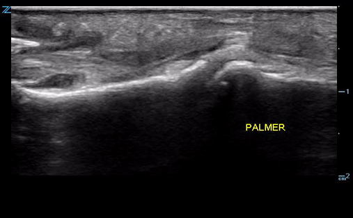
You attempt scalpel drainage (again using ultrasound) and only get sanguineous output, but on rescan you no longer see a distinct fluid collection in the soft tissue. The ortho PA attempts a large I&D of a presumed felon but only gets bloody output as well. In the mean time patient gets labs including ESR and CRP that are all normal.
Given diagnostic uncertainty, patient is admitted to ortho for observation overnight so she can be seen by the hand surgeon in the morning. She gets a dose of antibiotics but ultimately goes to the OR the following morning for exploration and washout. She is diagnosed with septic DIP joint, possible osteomyelitis and started on long term antibiotics even though all deep tissue cultures ended up being negative.
Still a bit of a mystery case to me. Probably the right choice to treat as infection, however given all labs, fluid and deep tissue cultures were negative, as well as the fact that no purulent discharge was ever found and patient had multiple bony fractures, I would still lean towards soft tissue swelling/ hematoma. Take away here is that ultrasound helped us differentiate between likely abscess / hematoma and FTS and was also helpful in guiding drainage.