
48 yom w flank pain
48 year old male presents with 2 days of intermittent severe left flank to LUQ abdominal pain unlike anything he has ever experienced before. Notes associated blood in the urine and nausea that occurs with episodes, no vomiting.
He has no history of prior kidney stones but you have high clinical suspicion while keeping your differential open.
Grab a curvilinear ultrasound probe, indicator to the head and take a look at both kidneys. Start with the non-affected side, then the affected side and don’t forget the bladder. Here is what we found.
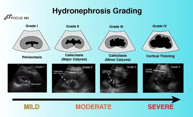
Moderate hydronephrosis demonstrated by large area of hypoechoic fluid in renal medulla without compression or thinning of cortex.
No uptake of color flow over hypoechoic areas indicate not vasculature but true hydronephrosis.
His pain was controlled and he was discharged home with urology follow up for renal stones.
Kidney stones affect 1 in 11 Americans. In 2014 Smith-Bindman et al. found that renal US and CT scan were statistically equivalent for diagnosing renal stones. This evidence was based on patients who were not thought to be high risk for serious alternative diagnoses such as aneurysms, bowel disorders or infections. They found that the use of US significantly reduced the exposure to radiation and decreased length of stays with no change in adverse outcomes.
Unilateral hydronephrosis on US in patients with a high index of suspicion for renal stone based on history is highly suggestive of obstructive renal stone. In males with bilateral hydronephrosis consider prostate problems. In females with bilateral hydro consider uterine problems.
Renal Ultrasound Made Easy: Step-By-Step Guide - POCUS 101
If you don’t see hydronephrosis remember that does not mean there is no stone, only that it is not obstructing.
You won’t always see a renal or ureteral stones but you can look for hyperechoic masses with a shadow similar to gallstones. Look for them in the kidney, the ureteral pelvic junction and the ureteral vesicular junction as they are trying to enter the bladder.
Another way to enhance your diagnosis is to try color doppler ultrasound and look for ”twinkling artifact” where irregular reflecting interfaces like stones or crystals will look like a mixture of red and blue pixels (see AlSaiady et al. article below).

Fig. 1. A. gray scale of renal stone. B. Color doppler with twinkle artifact. C. CT confirming renal stone.
1. AlSaiady M, Alqatie A, Almushayqih M. Twinkle artifact in renal ultrasound, is it a solid point for the diagnosis of renal stone in children? J Ultrason. 2021 Nov 29;21(87):e282-e285. doi: 10.15557/JoU.2021.0048. Epub 2021 Dec 15. PMID: 34970438; PMCID: PMC8678643.
2. Smith-Bindman R, Aubin C, Bailitz J, Bengiamin RN, Camargo CA Jr, Corbo J, Dean AJ, Goldstein RB, Griffey RT, Jay GD, Kang TL, Kriesel DR, Ma OJ, Mallin M, Manson W, Melnikow J, Miglioretti DL, Miller SK, Mills LD, Miner JR, Moghadassi M, Noble VE, Press GM, Stoller ML, Valencia VE, Wang J, Wang RC, Cummings SR. Ultrasonography versus computed tomography for suspected nephrolithiasis. N Engl J Med. 2014 Sep 18;371(12):1100-10. doi: 10.1056/NEJMoa1404446. PMID: 25229916.
3. Renal Ultrasound Made Easy: Step-By-Step Guide. Primary Authors: Jade Deschamps, Vi Dinh; Co-authors: Jessica Ahn, Satchel Genobaga, Annalise Lang, Victor Lee, Reed Krause, Devin Tooma, Seth White. Oversight, Review, and Final Edits by Vi Dinh (POCUS 101 Editor). Renal Ultrasound Made Easy: Step-By-Step Guide - POCUS 101